







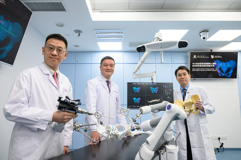
The Chinese University of Hong Kong has completed Hong Kong’s first robot-assisted pelvic fracture reduction and fixation surgeries, treating three patients at Prince of Wales Hospital since June.
The surgeries offer a glimpse of where medical robotics is heading, with imaging, navigation, and robotic movement increasingly working together while surgeons remain in control.
How the robotic system works
According to CUHK’s Faculty of Medicine, surgeons use 3D CT scans to plan how the fractured bone should be realigned and fixed before the operation. During surgery, real-time 3D navigation guides an active robotic arm as the fracture is repositioned and stabilized.
CUHK reported blood loss of 10 to 20 mL in its first three cases, compared with as much as one liter during conventional open surgery. It also said the minimally invasive approach reduced incisions from more than 10 centimeters to under two centimeters.
The university says the system has National Medical Products Administration clearance for pelvic and long-bone fractures and is listed under Hong Kong’s Medical Device Administrative Control System. CUHK did not identify the manufacturer in its announcement.
CUHK estimates Hong Kong sees about 2,000 fragility pelvic fractures each year and cites a roughly 15% one-year mortality rate among these patients. Older patients may also have other medical conditions that make major open surgery harder to tolerate.
The milestone comes as medical robotics expands across the region. Chinese surgical robot exports rose 3.3-fold in the first half of 2026, while a separate Chinese robotic system recently cut brain-imaging procedure time by about 29% in a 50-patient study.
Surgical robots are taking on more precise tasks
The immediate future of robotic surgery is less about autonomous machines operating alone and more about giving robots tightly defined technical jobs that complement a surgeon’s judgment.
Pelvic fracture treatment is well suited to that approach. CT scans provide detailed anatomical landmarks that can be turned into a 3D plan. Navigation systems can track instruments against that plan, while robotic arms can reproduce calculated positions and movements with high precision.
Research is beginning to quantify those advantages. A multicenter randomized trial published this year compared robotic and manual closed reduction in 92 adults with displaced pelvic fractures.
Excellent or good reductions were achieved in 96% of the robotic group versus 48% of the manual group. Surgeons in the robotic group also had a median of zero fluoroscopy exposures, referring to the X-ray imaging used to guide procedures, compared with 38 in the manual group.
A 2025 review of robot-assisted pelvic fracture fixation found similar gains in accuracy and reduced use of fluoroscopy across earlier studies.
The broader direction is toward surgical systems that connect more of these steps. Imaging can inform a digital plan, navigation can track anatomy and instruments in real time, and robotic hardware can carry out precise movements under a surgeon’s supervision.
AI could eventually add another layer by helping identify anatomy, flag risks, or adjust plans as conditions change during an operation. The surgeon would still make the clinical decisions, but fewer technical steps would need to be performed entirely by hand.
CUHK’s first pelvic procedures fit that emerging model. The robot is not replacing the surgeon. It is converting medical images and a surgical plan into controlled physical movement, one of several areas where robotics is becoming part of the operating room rather than a separate piece of equipment.
Also read: Robotic heart surgery is also moving major procedures toward smaller incisions, with El Camino Health recently using the da Vinci 5 for minimally invasive coronary bypass surgery.